
VISUAL ARCHIVE / IMAGE STORY
A Developmental Opening
A developmental genetic alteration left much of the mouse cerebral cortex absent, creating room for transplanted human cortical organoids.
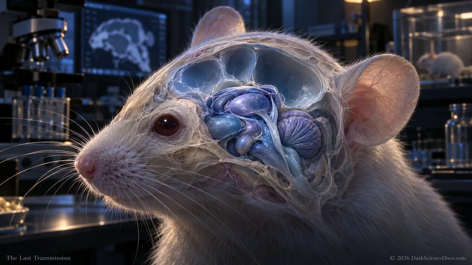
A developmental genetic alteration left much of the mouse cerebral cortex absent, creating room for transplanted human cortical organoids. The remaining brain structures provided a living environment in which the grafts could develop. Conceptual illustration.
THE OBSERVATION
What we can actually see.
At three months, graft tissue accounted for approximately 91.9 percent of the combined cortical tissue volume in the measured group of seven animals. That is a cortical comparison, not the fraction of an entire brain. Anatomical tracing and activity measurements supplied different kinds of evidence for integration. The article also describes above-chance alternation in a Y-maze among graft-bearing mice, unlike the ungrafted cortex-depleted comparison group.
THE ILLUSTRATION
What the artwork imagines.
A developmental genetic alteration left much of the mouse cerebral cortex absent, creating room for transplanted human cortical organoids. The remaining brain structures provided a living environment in which the grafts could develop. Conceptual illustration. The viewpoint, illumination, colors and simultaneous arrangement are artistic choices. This is a cinematic illustration, not a direct instrument image or a quantitative reconstruction of every feature.
FROM THE ILLUSTRATED ARTICLE
The story that inspired it.
Human-derived cortical tissue grew and connected inside mice engineered to develop without most of their cortex. The experiment opens a model of neural development while leaving the meaning of its behavioral results carefully bounded. This frame focuses on a developmental opening, within the complete illustrated article and its source-linked research context.
Read They Grew a Human Cortex Inside a Mouse. Then They Tested Its Memory.CHECK THE RECORD
The image interprets the research below. Follow the original work for the measurements and limits behind the scene.
Developmental xenocortication using human-derived organoids in mice Stanford Medicine research-team account, 2026-09-16 10.1038/s41467-022-35536-3 10.1016/j.stem.2023.01.004OTHER IMAGE STORIES
Keep looking.
 Human Cortical Tissue in a Mouse Model
 The Expanding Cortical Graft
 The Mark Shared by Titan and Pluto
EXPLORE IN 3D / ATLAS I-02
Look inside an illustrative cell
Compare cellular structures in an interactive model. This is a general cell explorer, not a reconstruction of the cortical-graft experiment.
Explore Cell in 3D