Cross the membrane, find the genome, and trace the cellular machinery. Choose a structure to reveal its story and the observations behind it.
INNER WORLD / 02 ANIMAL-TYPE CELL · SCHEMATIC SCALE
PLASMA MEMBRANE / CELL OVERVIEW
Choose a structure. Drag to rotate; pinch or use the zoom buttons.
FILE 01 / LIVING SYSTEMS
The Boundary
Plasma membrane / a selective interface
What makes a cell an inside?
The plasma membrane separates the interior from its surroundings and regulates exchange. This translucent shell keeps internal structures visible while giving the cell a defined edge. Its color and apparent thickness are artistic choices; this model does not resolve a lipid bilayer or membrane proteins.
How does a cell keep its genetic record accessible?
Most of this cell’s DNA is housed inside a membrane-bound nucleus. Pores in the nuclear envelope regulate traffic with the rest of the cell; the nucleolus is a region involved in making ribosomal components. The central sphere and luminous threads are a diagram of these relationships, not a mapped chromosome arrangement or a microscope image.
Why do these organelles have a history of their own?
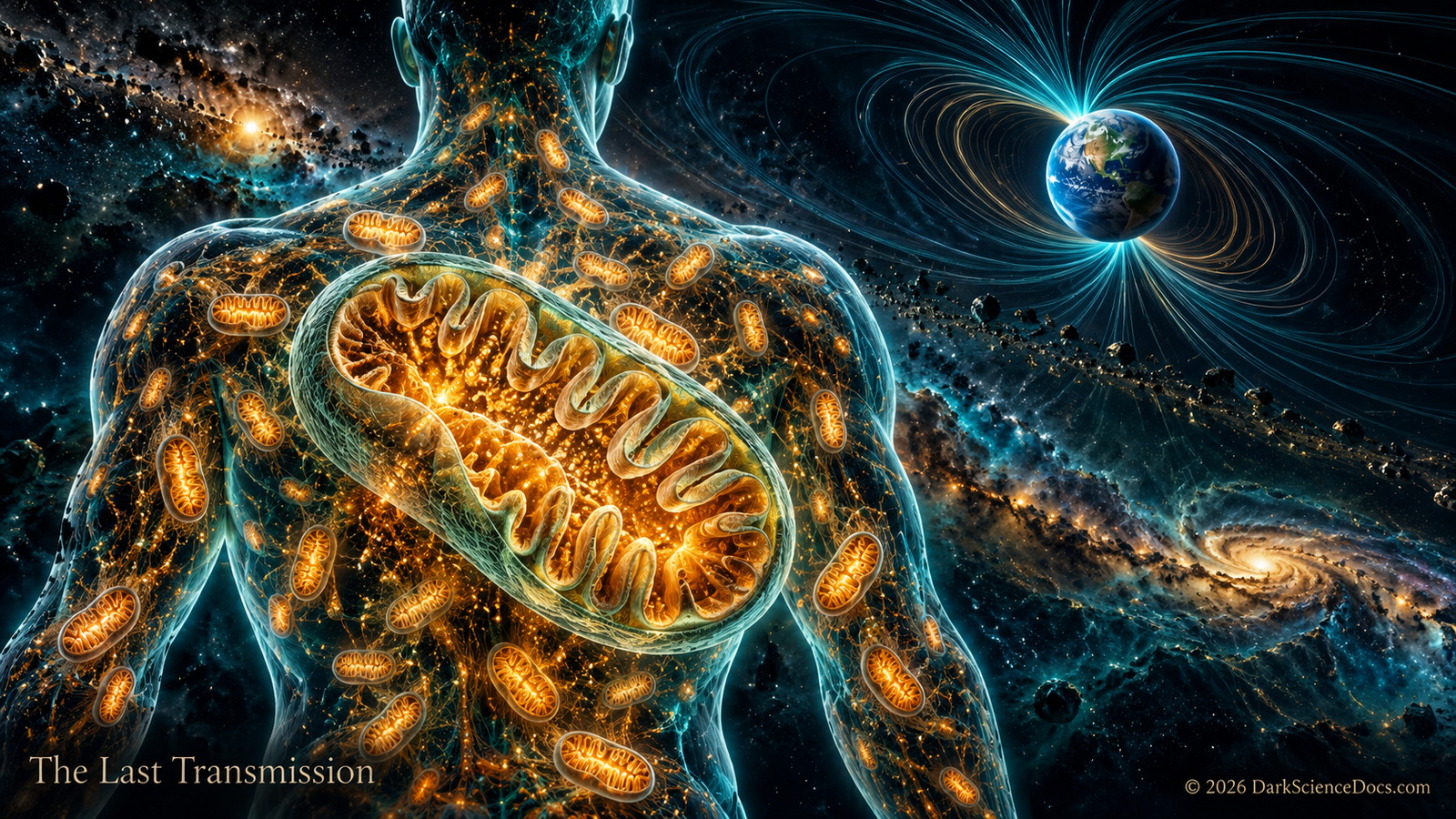
Mitochondria use chemical gradients across their folded inner membranes to help produce ATP. They also retain a small genome. Genetic and biochemical evidence supports a bacterial ancestry through endosymbiosis; the timing and sequence of the larger eukaryotic transition remain research questions. The four glowing bodies here stand in for a variable, often dynamic network, not a measured count or position.
The rough endoplasmic reticulum lies near and connects with the nuclear envelope. Ribosomes attached to it make proteins that enter the secretory pathway. In this scene, thin folded ribbons and pinpoint lights suggest the ER and ribosomes without claiming a faithful three-dimensional reconstruction of any one cell.
The Golgi receives proteins and lipids from the endoplasmic reticulum, modifies and sorts them, and helps dispatch them in vesicles. Its flattened stacks are recognizable in cell images, but the arcs and floating beads shown here are a simplified route map. Individual cargo molecules and their motion are not simulated.
Mitochondria point toward a bacterial ancestry within eukaryotes. Open the dedicated cutaway to examine their folded membrane, ATP-making machinery, and retained genetic material. The origin investigation follows a different, earlier question about the first free-living lineages.
This is a representative animal-type eukaryotic cell, drawn as a translucent teaching model. It is not a species-specific cell, a live simulation, or a photograph. Organelles vary between cell types and change shape and position over time. Their sizes, numbers, colors, and paths are arranged here for visibility; plant cells also have structures absent from this model, including chloroplasts and a cell wall.
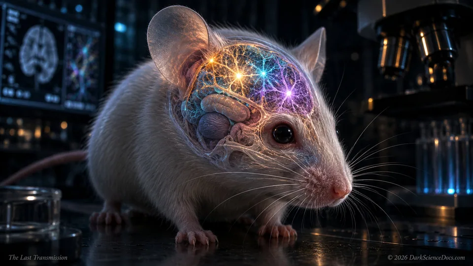
Scientists created mice whose cortical tissue was almost entirely derived from human cells. The tissue grew, formed connections, generated electrical activity, and revealed something extraordinary.
Compare black-hole horizons, calculate a signal’s travel time, and zoom from a cell toward an atom. Every tool shows its working and the assumptions behind its answer.
The cutaway organizes structures so their relationships can be explored. Real cells differ in shape, composition, and activity, and some structures are enlarged for visibility. Follow the reading links for the evidence behind a particular biological claim.
Ask yourself Which assumptions drive this result, and where would the model stop being useful?
THE SIGNAL CONTINUES
Some discoveries stay with you.
Get the illustrated investigations delivered to your inbox. Then follow the questions that refuse to leave.