
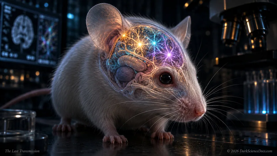
09 / Living systems / a developmental experiment
What a Human Cortical Graft Can Reveal Inside a Mouse
Human-derived cortical tissue grew and connected inside mice engineered to develop without most of their cortex. The experiment opens a model of neural development while leaving the meaning of its behavioral results carefully bounded.
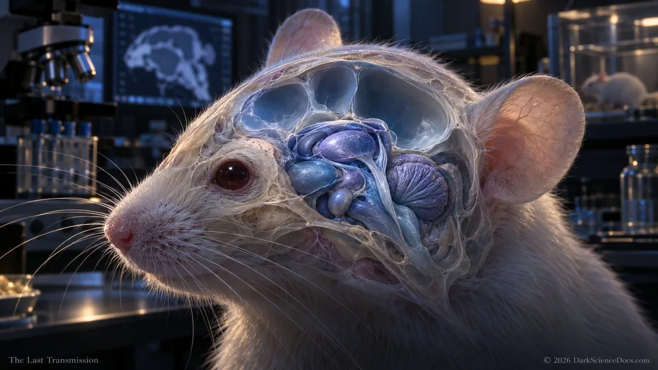
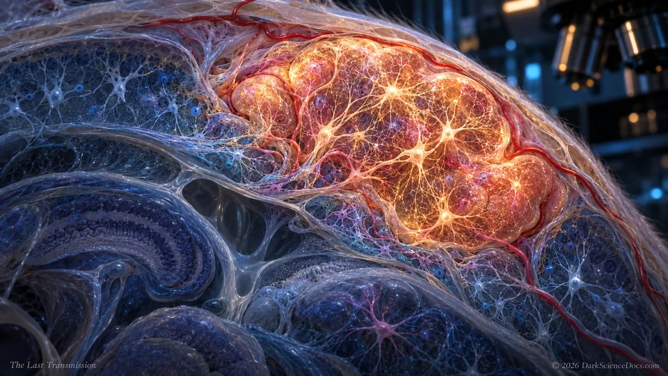
A nervous system is an environment as well as a collection of cells. Blood supply, incoming signals and neighboring structures influence how a transplanted organoid develops. In the reported experiment, newborn mice with a developmental genetic depletion of much of the cerebral cortex received human-derived cortical tissue. The grafts expanded, developed organized activity and formed projections into the host nervous system. The study therefore asks how human cells develop within a living animal, rather than whether an intact adult brain can simply be exchanged. Its biological scale is striking, but the measured denominator matters as much as the percentage.
The full investigation continues with the evidence, illustrations, and sources at The Last Transmission on Substack.
Continue reading on SubstackTHE EVIDENCE / THE OPEN QUESTION
What we know.
At three months, graft tissue accounted for approximately 91.9 percent of the combined cortical tissue volume in the measured group of seven animals. That is a cortical comparison, not the fraction of an entire brain. Anatomical tracing and activity measurements supplied different kinds of evidence for integration. The article also describes above-chance alternation in a Y-maze among graft-bearing mice, unlike the ungrafted cortex-depleted comparison group.
What remains open.
The experiment did not transfer a person’s memories or establish human consciousness in a mouse. Above-chance behavior does not by itself show which circuit was necessary for the task. Remaining host structures, developmental differences and the graft’s precise contribution require targeted comparisons. Long anatomical projections likewise cannot independently establish direct behavioral control.
Why it matters.
A better disease model can reveal how human neural tissue develops, responds to injury and interacts with a wider circuit. Establishing a causal behavioral contribution would require selective interventions alongside careful controls. That work is valuable without converting a developmental model into a claim about identity, human-level thought or an established treatment.
GO DEEPER / EVIDENCE FILE
What Did Human Cortical Grafts in Mice Actually Demonstrate?
Separate the developmental model, the cortical-volume denominator, neural integration and the limited behavioral inference in a human-organoid mouse experiment.
FOLLOW THE EVIDENCE
Research behind the field note.
EXPLORE IN 3D / ATLAS I-02
Look inside an illustrative cell
Compare cellular structures in an interactive model. This is a general cell explorer, not a reconstruction of the cortical-graft experiment.
Explore Cell in 3DVISUAL EVIDENCE
Inside the investigation.
FOLLOW THE THREAD
Related signals.
They Found Plutonium at the Bottom of the Pacific. The Explosion That Made It May Be Over 95 Million Years Old.
Deep beneath the ocean, scientists discovered radioactive debris from beyond Earth. The most disturbing clue was an element they could not find.

The Blows That Nearly Killed Earth May Have Helped Bring It to Life
A model identifies a window for life’s chemistry 4.33 billion years ago. The unsettling question is whether Earth was the only world to take advantage of it.

Before the Forests, There Were Hunters
Arachnids were hunting on land long before the first forests appeared. The fossils behind their story reveal an ancient invasion and remarkable inventions.