FILE 01 / PROKARYOTIC CELL
The Envelope
Cell wall / plasma membrane
What holds this cell together?
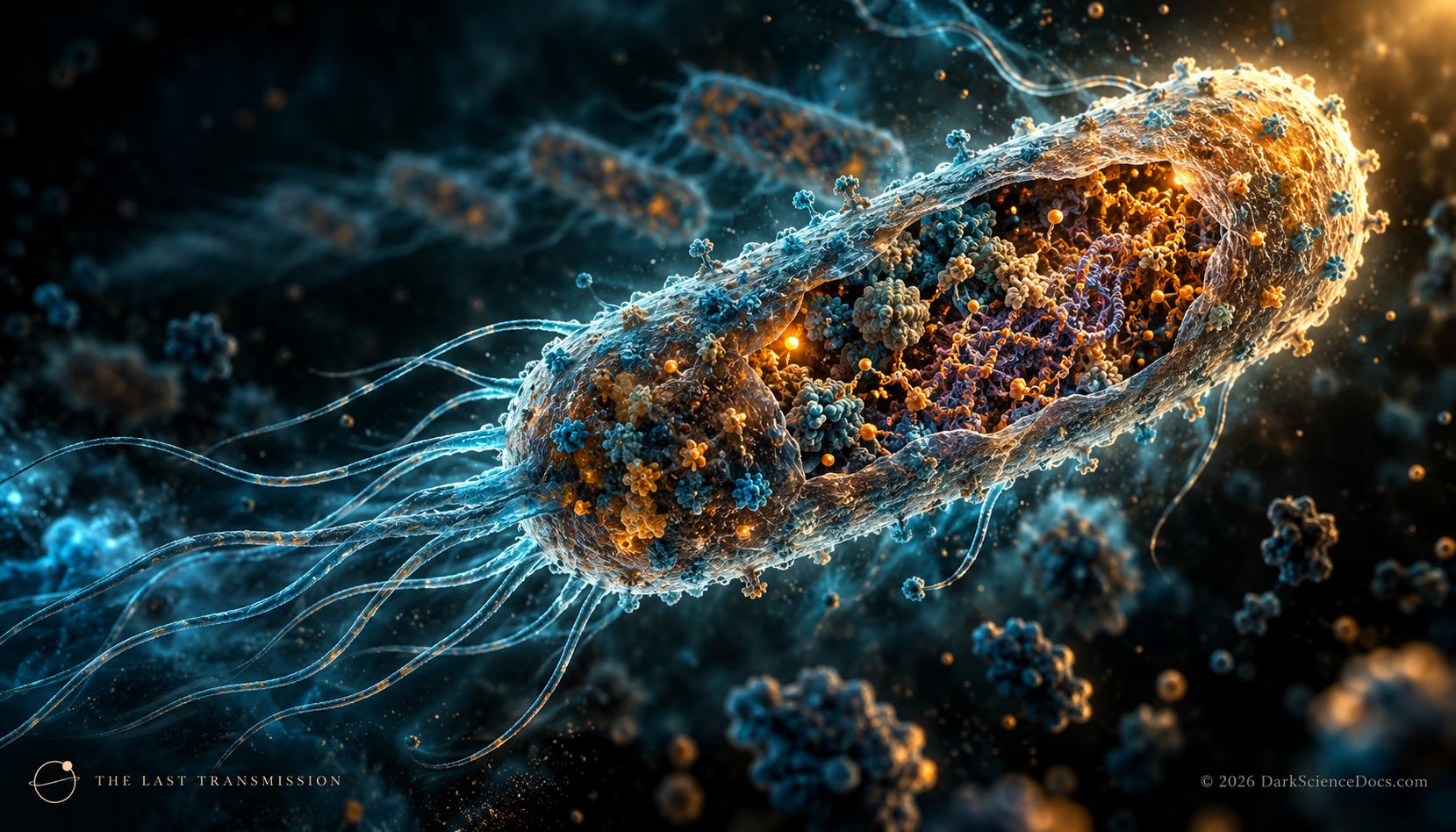
This illustration shows one bacterial plasma membrane enclosed by a peptidoglycan wall. The membrane controls exchange and the wall helps the cell resist pressure. Bacterial envelopes vary: some have an additional outer membrane, some have capsules, and a few lack a wall. Archaea are also prokaryotes, but their membrane lipids and wall chemistry differ. The two glowing outlines here are simplified layers, not a resolved molecular cross-section.
FILE 02 / PROKARYOTIC CELL
The Nucleoid
Chromosomal DNA / no nuclear envelope
Where is the genome when there is no nucleus?
Bacterial chromosomal DNA occupies a region called the nucleoid, which is not enclosed by a nuclear membrane. Many bacteria have a circular chromosome, though that is not universal. The folded luminous strand represents organized DNA without reproducing the shape, copy number, or gene order of any species. The open cytoplasm is a real contrast with the membrane-bound nucleus in the animal-type cell map.
FILE 03 / PROKARYOTIC CELL
The Ribosomes
Protein synthesis / distributed through cytoplasm
How does the cell turn genetic instructions into proteins?
Ribosomes assemble proteins from information carried by RNA. This bacterial cytoplasm contains many ribosomes, but the pinpoint lights are only a sparse visual sample. Their count, location, and scale do not represent a measured cell. A bacterium does not need a membrane-bound nucleus for this molecular work to happen.
FILE 04 / PROKARYOTIC CELL
Energy at the Membrane
Electron transport / proton gradient / ATP
What does energy production look like without mitochondria?
In bacteria that use an electron transport chain, proteins in the plasma membrane can pump hydrogen ions across it and help establish a gradient. ATP synthase can use the return flow to make ATP. Metabolism is diverse across bacteria, so this pathway is an example, not the only way a prokaryote lives. Blue points mark the outer side of the membrane schematically; they are not a live proton count or animation.
FILE 05 / PROKARYOTIC CELL
A Possible Plasmid
Extra DNA / present in some bacteria
What other genetic instructions can a cell carry?
Some bacteria carry plasmids: DNA molecules separate from their chromosome. Certain plasmids can move between cells and carry traits such as antibiotic resistance. The small loop here marks a possibility, not a universal component. It does not depict a particular plasmid, a transfer event, or a measured genome.
FILE 06 / PROKARYOTIC CELL
A Possible Flagellum
Optional motility structure
How might a bacterium move?
Some bacteria swim with one or more flagella, while many do not have one. A bacterial flagellum is a rotating motility structure. The long curved filament shown here is stationary and enlarged for visibility; it does not reproduce a species-specific motor, number of flagella, or swimming behavior.